MeVisLab provides many powerful tools and features for visualizing your project from medical to industrial imaging.
Today we would like to highlight the MeVisLab Path Tracer which was used by biologists at the University of Helsinki to develop a series of networks for implementing photorealistic renderings, capturing tissue-level to whole-organism details across diverse living organisms.
The powerful combination of SmARTR networks with the MeVisLab Path Tracer is particularly impressive when directly comparing real specimens with their rendered counterparts, as in seen below in panel one.
The published results are available here: “The SmARTR Pipeline: a modular workflow for the cinematic rendering of 3D scientific imaging data”.
Panel 1:
(Top row): Images of a live specimen of the lizard Anolis carolinensis (left) and the dissected heart from the bearded dragon lizard, Pogona vitticeps (right). (Bottom row): 3D renderings of the heart of Pogona vitticeps (top left), the sectioned head of Anolis carolinensis highlighting the skull and brain (top right), and an intact specimen of Anolis carolinensis (bottom). SmARTR Networks Used: SmARTR_Multi-Independent_Volume network for the Pogona vitticeps heart, SmARTR_Advanced_Multi-Volume network for the sectioned head, and SmARTR_Pattern_Drawing for the intact Anolis carolinensis specimen.
Panel 2:
(Top row): Renderings of the head of the snake Cerastes cerastes—intact (left) and sectioned to highlight the venom system (right). (Bottom row): Renderings of the anterior part of the hillstream loach, Gastromyzon zebrinus—sectioned to highlight the skeleton, brain, and gills (left) and intact (right). SmARTR networks used: SmARTR_Pattern_Drawing for the intact specimens; SmARTR_Advanced_Multi-Volume network for the cutaway views.

Panel 3:
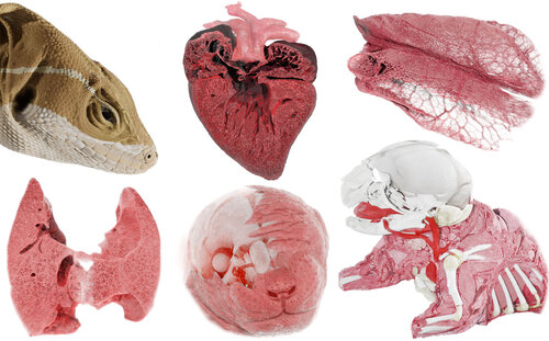
(Top row): Renderings of the head of the lizard Basiliscus vittatus (left), and the heart (middle) and lungs (right) of the lizard Pogona vitticeps, both sectioned to reveal internal structures. (Bottom row): Renderings of the lungs (left), head (middle), and trunk (right) of the house mouse, Mus musculus. SmARTR networks used: SmARTR_Pattern_Drawing for the intact specimen; SmARTR_Nested_Multi-Volume network for the mouse head, lungs, and trunk, and lizard lungs; SmARTR_Advanced_Multi-Volume network for the cutaway view of the lizard heart.

Panel 4:
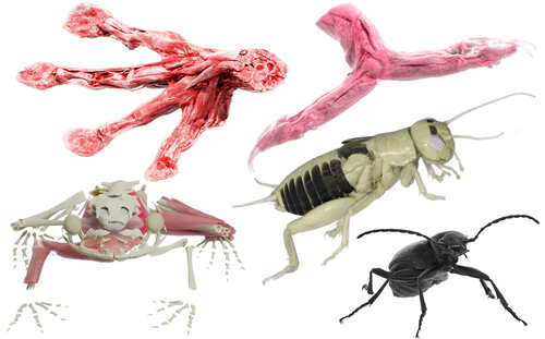
(Top row): Forelimbs of the tree frog Agalychnis callidryas (left) and the dwarf chameleon Chamaeleo calyptratus (right). (Bottom row): Rendering of the skeleton and internal organs of the tree frog Agalychnis callidryas (left), the intact body of the cricket Gryllus bimaculatus (middle), and the ground beetle Pterostichus oblongopunctatus (right). SmARTR networks used: SmARTR_Single-Volume for both species' forelimbs and the beetle rendering; SmARTR_Multi-Independent_Volume network for the frog's skeleton and organs; SmARTR_Pattern_Drawing for the intact cricket specimen.

Panel 5:
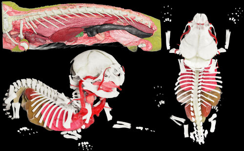
(Top row): Cutaway view of the trunk of the dwarf chameleon Chamaeleo calyptratus, highlighting skeletal elements and internal organs. (Bottom row): Skeletal elements and internal organs of the house mouse Mus musculus in fronto-lateral view (left) and dorsal view (right). SmARTR networks used: SmARTR_Nested_Multi-Volume network for the chameleon's trunk cutaway view; SmARTR_Multi-Independent_Volume network for the mouse's skeletal